Article in association with Keeler UK and AOS – Advanced Ophthalmic Systems
![]()

The past 10 or 12 years have seen huge advancements in ocular imaging. Colour fundus cameras, OCT for both posterior and anterior segments, wide field retinal imaging and other techniques such as auto-fluorescence are just some of the systems gracing optometric practice nowadays.
Only 20 years ago clinical examination of the eye mainly consisted of a look at the anterior segment with the slit-lamp, followed by fundal examination with the direct ophthalmoscope. Even dilation and use of a volk lens would have been a technique carried out in very small minority of cases. This revolution in imaging has been a tremendous step forward for our profession. It allows us to detect and diagnose conditions more accurately, provide a much higher level of information to ophthalmology colleagues when referring, help educate patients on their condition, and enhances our ability to monitor our patients.
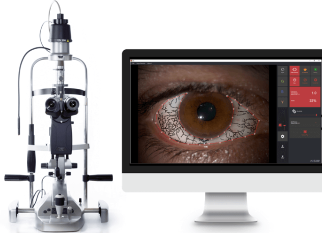
This article brings to you the latest such step forward in ocular imaging. This time it is concentrated on the anterior segment of the eye. AOS (Advanced Ophthalmic Systems) along with Keeler have developed and released ‘AOS Anterior’ – a software package designed to work with any existing anterior segment camera, which helps to grade clinical signs to a much more accurate and repeatable degree than before.

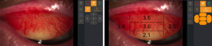
This system allows the user to photograph the anterior segment and accurately measure and grade Bulbar Redness, Lid Redness and Corneal Staining. Up to now these features have been graded using clinical grading scales. While very useful, these can’t give an exact quantification of the clinical finding in the same way that this system measures, and are always going to be open to subjective interpretation of different users. A study by London City University found the system to be more repeatable and reliable than conventional grading scales.
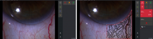
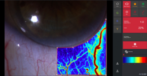
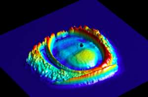
The AOS system grading of conjunctival and bulbar redness extracts the linear patterns of the blood vessels and then quantifies this both in terms of a grade from 0 to 4 and as a percentage of the overall area covered by this injection. The image of the injected conjunctiva can also be displayed as a heat map, which is interesting in terms of giving a sense of the underlying inflammation. This also provides a great visual for the patient.
When an area of the cornea is selected for punctate staining, again the system recognises each point of staining and counts these to give a definitive measure.
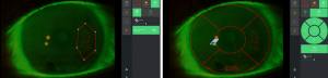
The software works in conjunction with the existing slit-lamp and anterior camera systems. The area of interest is photographed and the software automatically grades this and gives a score, whether it is an area of conjunctival redness or and area of corneal staining for example. The user has the ability to map the exact area that they want graded with a drawing tool.
This ability to accurately grade and monitor would be incredibly useful in certain patient groups such as contact lenses, dry eye evaluation, red eye and blepharitis. In all of these groups it could be a great way to illustrate to patients what is happening with their ocular surface and to help to get them onboard with your treatment, be it importance of ocular lubrication, lid cleaning or with the choice of a new contact lens material or regime.
It is not often I get the opportunity to meet the person who develops a new piece of kit. In this case I had a great conversation with Dr Eduardo Mangieiri, who developed this software. Eduardo had been involved in imaging and grading clinical signs and features for other medical specialties such as cardiology, before being involved in grading systems for retinal vasculature. For him, it seemed an obvious leap to apply his expertise to developing a system which can make our assessment and monitoring of anterior segment changes much more accurate. We chatted about other possibilities of this technology and he is currently working on applying this to the grading of meibomian gland dysfunction. There are also plans to incorporate a measuring tool in the software when AOS v2 arrives this year, which could be invaluable for measuring lesions such as ulcers and abrasions and monitoring how they heal. This next generation of the software will also allow 3-d imaging of fluorescein patterns on contact lenses, which may make for more accurate fitting – particularly in more complex cases such as keratoconus.
As updates are new features are developed, these can be incorporated into systems for existing users, so this product is an ongoing package for the user. I strongly suspect over the next few years, this type of enhanced means of detecting and recording anterior signs will become common place in practice, in a similar way to how we image the fundus. As electronic patient records are also slowly becoming the default, this type of technology should integrate easily with the patients’ records and provide an excellent way for practitioners to monitor patient progress.
This will certainly be an interesting area of advancement to watch in the coming years.
Thank you for reading.
Stanley Keys.
![]()
For more information please visit follow the links for Keeler or AOS above, or you can contact Keeler UK directly:
Keeler Ltd, Clewer Hill Rd, Windsor, SL4 4AA Tel 01753 857177
Or Contact Brian Moran of Keeler brianm@keeler.co.uk Tel 07836248139